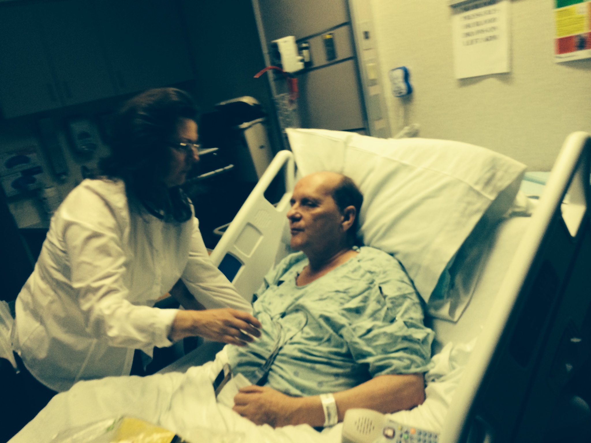
1. The Emergency Room Dilemma
The stroke clock is a merciless metronome. In emergency departments across the globe, the arrival of a patient with a sudden facial droop, slurred speech, and a limp arm triggers a high-velocity protocol. The “time is brain” mantra dictates every movement: the frantic wheeling of the gurney to CT, the rapid-fire neurologic checks, and the heavy decision to administer “clot-busting” thrombolytics. For the patient, it is a moment of pure terror—a sudden loss of self. For the clinician, it is a race to save millions of neurons from ischemia.
However, neurology is increasingly confronting a diagnostic shadow. We are discovering that the brain possesses a long, haunting memory of its past injuries. Sometimes, the symptoms that look like a brand-new medical emergency are actually “ghosts” of an old injury, unmasked by a completely unrelated stressor elsewhere in the body. As we refine our diagnostic tools, we are finding that every neurological deficit need not be a new stroke.
2. The 60% Statistical Shock: When a Stroke Isn’t a Stroke
While speed remains the priority in the ER, recent data reveals how often clinical appearances can be deceiving. A case-control study published in the Journal of Community Hospital Internal Medicine Perspectives (JCHIMP) examined patients who received alteplase (tPA), the gold-standard treatment for acute stroke. Among the subset of patients who showed normal initial vascular imaging, a staggering 60% were eventually diagnosed as “stroke mimics.”
The statistical markers for these mimics are profound. According to the study, stroke mimics had 10.1 times higher odds of having a history of prior stroke and 12.9 times higher odds of a history of migraine than those suffering a true vascular event. These mimics often arise from:
- Seizures: Specifically the postictal state known as Todd’s paralysis.
- Migraines: Particularly complex episodes featuring aura or sensory deficits.
- Conversion Disorder: Functional symptoms often precipitated by acute psychological stress.
- Metabolic Insults: Severe electrolyte imbalances like hyponatremia or toxic encephalopathy.
Identifying these mimics early is a matter of both patient safety and systemic efficiency. A typical stroke hospitalization can exceed $20,000, while a targeted MRI workup costs a fraction of that amount. Distinguishing a mimic prevents unnecessary, expensive interventions and saves the patient from the potential side effects of unneeded high-risk medications.
3. Post-Stroke Recrudescence (PSR): The “Anamnestic” Return
Perhaps the most perplexing of these mimics is Post-Stroke Recrudescence (PSR). Rather than a new injury, PSR is the transient reemergence of neurological deficits from a previous “index” stroke. PSR typically lies in wait for nearly four years—averaging an onset of 3.9 years post-injury—before suddenly flaring up.
This is an anamnestic process—a physiological “recall.” The brain isn’t bleeding or blocked; it is simply decompensating. The symptoms are usually abrupt yet mild, typically affecting the motor-sensory or language pathways that were damaged years prior. To help clinicians differentiate this “ghost” from a new attack, the following framework is utilized:
The 5-Point Diagnostic Framework for PSR
- Transient worsening of residual post-stroke focal neurological deficits.
- Evidence of a chronic stroke on brain imaging that matches the clinical distribution of symptoms.
- Absence of acute lesions on modern MRI-DWI (diffusion-weighted imaging).
- Clinical ischemia deemed unlikely (symptoms typically last >1 hour without new imaging evidence; absence of high-grade arterial stenosis).
- Exclusion of seizures through clinical observation or EEG.
4. The Unexpected Triggers: From UTIs to Sleep Deprivation
How does a brain “remember” an injury so vividly? The answer lies in a loss of “physiological reserve.” While the brain can reorganize and bypass a site of injury through neuroplasticity, these new pathways have a lower threshold for failure. When the body is stressed, these compensatory networks are the first to buckle.
The triggers for this reactivation are often surprisingly mundane:
- Infections: Systemic stressors, specifically Urinary Tract Infections (UTIs) and Pneumonia, are the primary culprits.
- Metabolic Shifts: Significant drops in blood pressure (hypotension) or low sodium (hyponatremia).
- The Medicine Cabinet: Sedatives, benzodiazepines, or opioids can destabilize the synaptic networks maintaining the brain’s recovery.
- Lifestyle Stress: Acute psychological trauma and severe insomnia.
A leading theory for this involves “Immune Priming.” During the initial stroke, the breakdown of the blood-brain barrier exposes the immune system to Myelin Basic Protein (MBP)—a “nonspecific brain self-antigen” normally hidden from view. This exposure primes the immune system’s memory. Years later, an unrelated infection triggers a cytokine cascade that reactivates these primed cells. They home in on the old, structurally abnormal stroke site, temporarily disrupting its function.
Dr. Aneesh B. Singhal, a vice chair of neurology at Massachusetts General Hospital, notes that physicians must be vigilant for these triggers:
“If a patient re-presents with a worsening of their original symptoms, [physicians] should consider the diagnosis of recrudescence rather than… a new stroke, and their workup would include urinalysis, [chest X-rays for] pneumonia, and cultures.”
5. The Case of the Ramsay Hunt “Mimic”
The reality of PSR is illustrated by the case of a 70-year-old woman admitted for what appeared to be an acute posterior circulation stroke. She presented with walking imbalance (ataxia) and facial paralysis. Her medical history was complex: she suffered from diabetes and hypertension, and six years prior, she had been hospitalized for diabetic cranial neuropathythat left her with an abduction deficit in her right eye.
At the time of admission, she was also battling Ramsay Hunt Syndrome (shingles reactivation). MRI imaging showed no new acute injury, but it did reveal the “ghost” of a chronic infarction in her pons (the brainstem). The shingles infection had acted as a systemic stressor, unmasking the vulnerabilities of her previous pons injury and her history of cranial neuropathy. Once her shingles was treated with antivirals and steroids, her “stroke” symptoms completely regressed, proving the cause was systemic, not vascular.
6. Better Safe Than Sorry: The Thrombolytic Safety Net
Despite the frequency of mimics like PSR, the medical community maintains a “treat first” stance within the 4.5-hour therapeutic window. The stakes of missing a real, treatable stroke are simply too high to wait for an MRI to confirm a mimic.
Fortunately, the safety of “accidentally” treating a mimic is remarkably high. Research confirms that when PSR patients or other mimics are given tPA (alteplase), the rate of complications—including symptomatic intracerebral hemorrhage—is exceptionally low. While doctors strive for diagnostic precision, the “stroke clock” ensures that aggressive, early treatment remains the safest default in the face of uncertainty.
7. Conclusion: The Vulnerable Brain’s Memory
Post-Stroke Recrudescence is far more common than previously recognized, representing approximately 10% of all presentations for transient neurological attacks. It serves as a stark reminder that the brain is a resilient but delicate organ, capable of harboring “shadows” of past trauma for decades.
If our brains can “remember” injuries so vividly that a simple UTI or a night of poor sleep can unmask them years later, it suggests that recovery is not a static destination, but a state of active, fragile compensation. We rely on a hidden “physiological reserve” every day—a backup system that keeps us functioning until a systemic insult pushes us over the edge.
As we look forward, we must ask: if the brain’s recovery is so contingent on our total systemic health, what other ghosts of past injuries are we currently compensating for, and how can we better protect the fragile balance of our neural reserves?